alb3806807
Brain, Anatomical Illustration, 1802
| Teilen |
|---|
 Pinterest Pinterest |
 Twitter Twitter |
 Facebook Facebook |
 Link kopieren Link kopieren |
 Email Email |
|
Zu einem anderen Lightbox hinzufügen |
|
Zu einem anderen Lightbox hinzufügen |


Haben Sie bereits ein Konto? Anmelden
Sie haben kein Konto? Registrieren
Dieses Bild kaufen

Titel:
Brain, Anatomical Illustration, 1802
Untertitel:
Siehe automatische Übersetzung
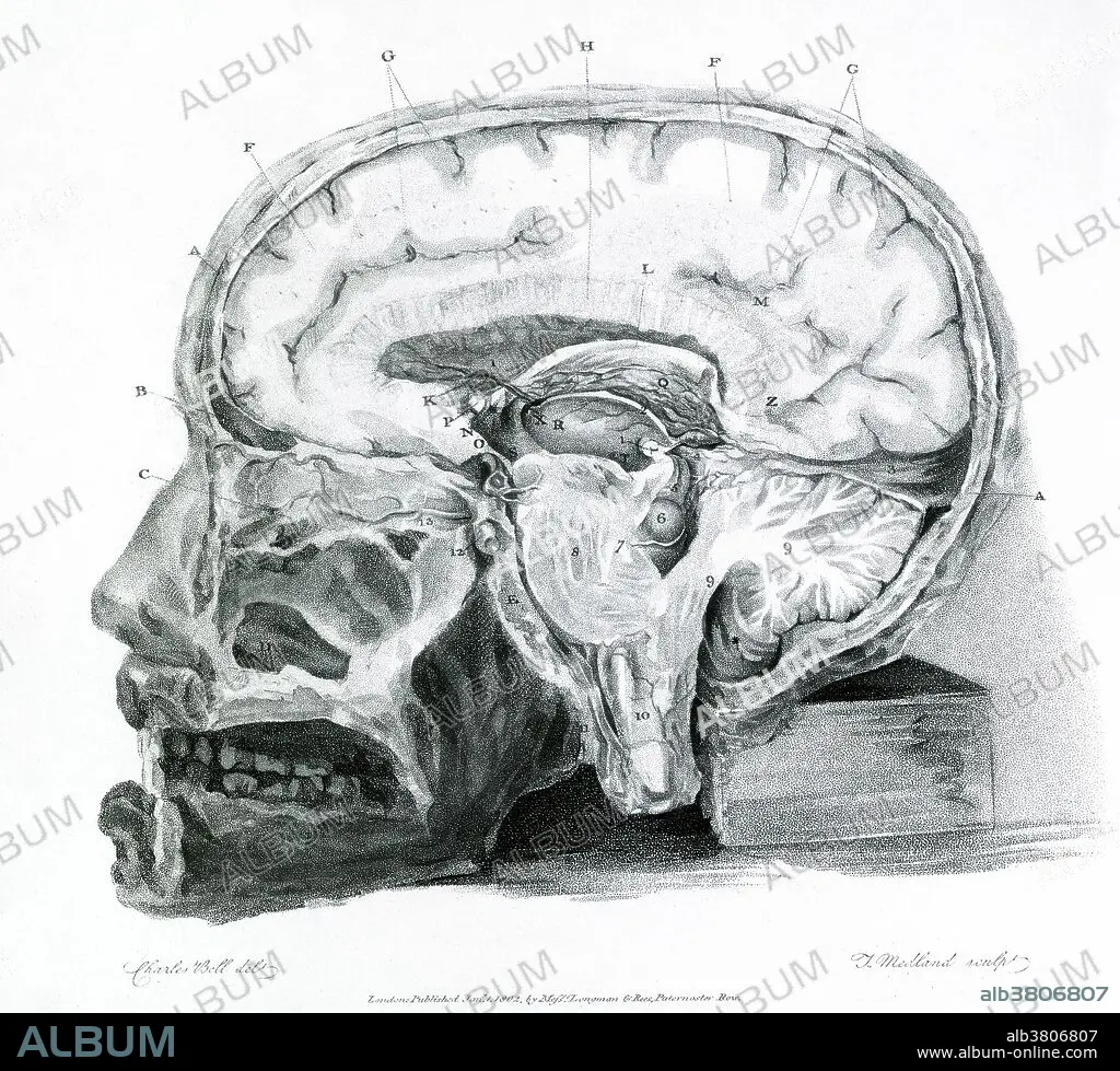
Historical illustration showing a section of the brain. From "The Anatomy of the Brain Explained in a Series of Engravings" by Sir Charles Bell, 1802. Sir Charles Bell (1774-1842) worked mainly on corpses, but he did conduct some neurological experiments on living animals, cutting or stimulating nerves to determine the localization of brain function: he could see no other means of demonstrating his belief in the differential function of the cerebrum and cerebellum, based on his work as a dissector. He established the basic distinction between anterior and posterior roots of the spinal nerves, which were later shown to govern movement and sensation respectively.
Bildnachweis:
Album / Science Source / Wellcome Images
Freigaben (Releases):
Model: Nein - Eigentum: Nein
Rechtefragen?
Rechtefragen?
Bildgröße:
3554 x 3214 px | 32.7 MB
Druckgröße:
30.1 x 27.2 cm | 11.8 x 10.7 in (300 dpi)
Schlüsselwörter:
