alb10687860
Normal coronal and cross-sections of brain, illustration
| Teilen |
|---|
 Pinterest Pinterest |
 Twitter Twitter |
 Facebook Facebook |
 Link kopieren Link kopieren |
 Email Email |
|
Zu einem anderen Lightbox hinzufügen |
|
Zu einem anderen Lightbox hinzufügen |


Haben Sie bereits ein Konto? Anmelden
Sie haben kein Konto? Registrieren
Dieses Bild kaufen

Titel:
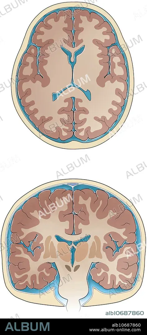
Normal coronal and cross-sections of brain, illustration
Untertitel:
Siehe automatische Übersetzung
Normal coronal and cross-sections of brain, illustration. Slices through the normal brain and skull showing ventricles, white matter, grey matter, basal ganglia, thalamus sulci and gyri, cerebrospinal fluid and skull.
Bildnachweis:
Album / Science Source / Sue Seif
Freigaben (Releases):
Model: Nein - Eigentum: Nein
Rechtefragen?
Rechtefragen?
Bildgröße:
2841 x 6150 px | 50.0 MB
Druckgröße:
24.1 x 52.1 cm | 9.5 x 20.5 in (300 dpi)
Schlüsselwörter:
ANATOMIE • ANATOMIE: SCHAEDEL • CRANIUM • GRAU • ILLUSTRATION • ILLUSTRATIONS • SCHAEDEL • SCHAEDEL, ANATOMIE • SCHÄDEL • TOTENKOPF • ZEICHNUNGEN
