alb10620608
ARACHNOID CYST
| Teilen |
|---|
 Pinterest Pinterest |
 Twitter Twitter |
 Facebook Facebook |
 Link kopieren Link kopieren |
 Email Email |
|
Zu einem anderen Lightbox hinzufügen |
|
Zu einem anderen Lightbox hinzufügen |


Haben Sie bereits ein Konto? Anmelden
Sie haben kein Konto? Registrieren
Dieses Bild kaufen

Titel:
ARACHNOID CYST
Untertitel:
Siehe automatische Übersetzung
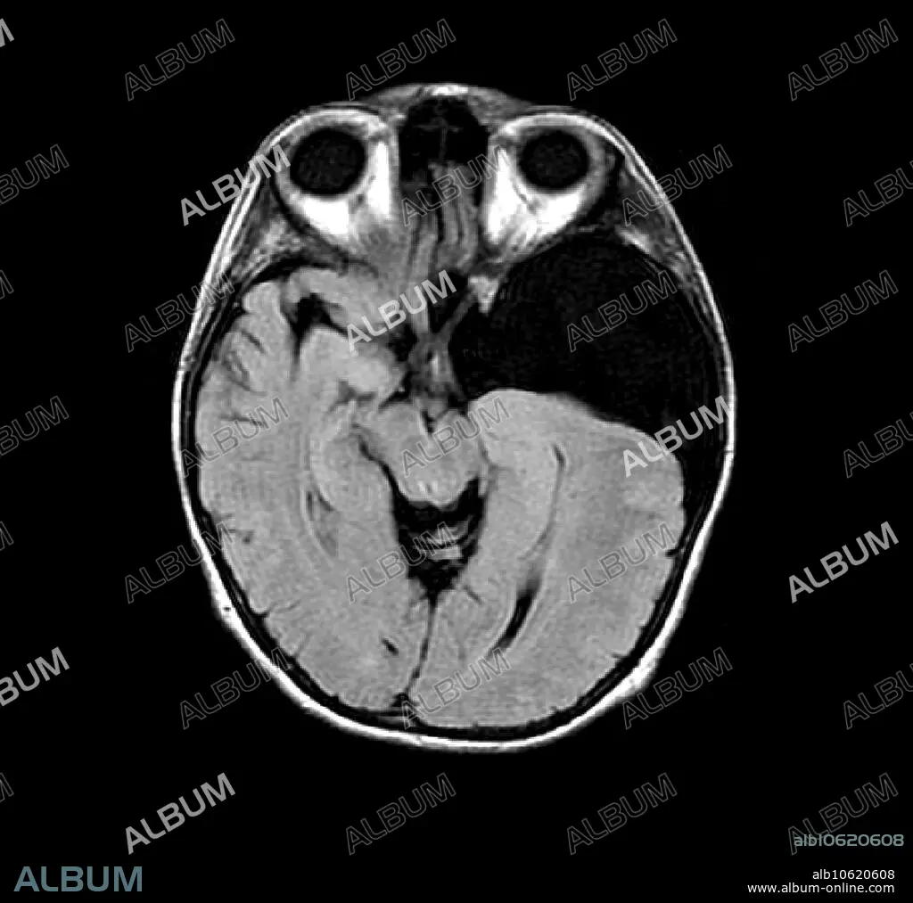
This axial (cross section) MRI image of the brain shows a large left middle cranial fossa arachnoid cyst (at right, dark). This is an abnormal collection of CSF (cerebral spinal fluid) which is walled off and acts like a mass. It is benign but can cause a variety of symptoms including seizures.
Bildnachweis:
Album / Science Source / LIVING ART ENTERPRISES, LLC
Freigaben (Releases):
Model: Nein - Eigentum: Nein
Rechtefragen?
Rechtefragen?
Bildgröße:
3842 x 3600 px | 39.6 MB
Druckgröße:
32.5 x 30.5 cm | 12.8 x 12.0 in (300 dpi)
Schlüsselwörter:
ANATOMIE • ANATOMIE: SCHAEDEL • BESCHLAGNAHME • CRANIUM • KLANG • SCHAEDEL • SCHAEDEL, ANATOMIE • SCHÄDEL • TOTENKOPF • UNORDNUNG
