alb10679191
cervical spine
| Teilen |
|---|
 Pinterest Pinterest |
 Twitter Twitter |
 Facebook Facebook |
 Link kopieren Link kopieren |
 Email Email |
|
Zu einem anderen Lightbox hinzufügen |
|
Zu einem anderen Lightbox hinzufügen |


Haben Sie bereits ein Konto? Anmelden
Sie haben kein Konto? Registrieren
Dieses Bild kaufen

Titel:
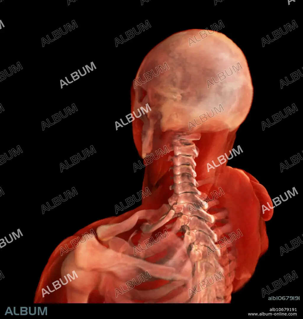
cervical spine
Untertitel:
Siehe automatische Übersetzung
3D visualization based on scanned human data of posterolateral view of the cervical spine. The cervical spine consist of 7 vertebrae - C1-C7. The movements of the cervical spine are flexion and extension of the head which predominantly take place between the first cervical spine and the occipital bone of the skull. Rotation of the head occurs entirely at the joint between the first and second cervical vertebrae, the atlanto-axial joint.
Bildnachweis:
Album / Science Source / ANATOMICAL TRAVELOGUE
Freigaben (Releases):
Model: Nein - Eigentum: Nein
Rechtefragen?
Rechtefragen?
Bildgröße:
2000 x 2000 px | 11.4 MB
Druckgröße:
16.9 x 16.9 cm | 6.7 x 6.7 in (300 dpi)
Schlüsselwörter:
ANATOMIE • ANATOMIE: KNOCHEN • ANATOMIE: SCHAEDEL • CRANIUM • DREI • GEGEND • HALS • KNOCHEN • MENSCH (MENSCHEN) • MENSCH • MENSCHLICH • NACKEN • SCHAEDEL • SCHAEDEL, ANATOMIE • SCHLUESSELBEIN • SCHLÜSSELBEIN • SCHÄDEL • SKAPULER • TOTENKOPF
