alb10656953
Lamina Externa, TEM
| Teilen |
|---|
 Pinterest Pinterest |
 Twitter Twitter |
 Facebook Facebook |
 Link kopieren Link kopieren |
 Email Email |
|
Zu einem anderen Lightbox hinzufügen |
|
Zu einem anderen Lightbox hinzufügen |


Haben Sie bereits ein Konto? Anmelden
Sie haben kein Konto? Registrieren
Dieses Bild kaufen

Titel:
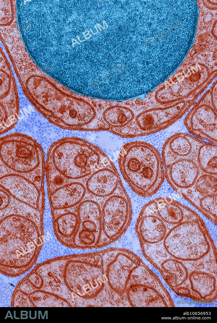
Lamina Externa, TEM
Untertitel:
Siehe automatische Übersetzung
Color enhanced transmission electron micrograph of a nerve in cross-section, showing groups of unmyelinated axons enclosed in deeply invaginated recesses in the surface of the Schwann cells. Around each Schwann cell is a thin, continuous lamina externa, especially visible at the arrows.
Bildnachweis:
Album / Science Source / DON W. FAWCETT
Freigaben (Releases):
Model: Nein - Eigentum: Nein
Rechtefragen?
Rechtefragen?
Bildgröße:
3490 x 4939 px | 49.3 MB
Druckgröße:
29.5 x 41.8 cm | 11.6 x 16.5 in (300 dpi)
Schlüsselwörter:
