alb10612319
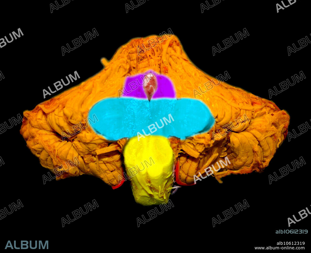
Normal Brainstem and Cerebellum

|
Zu einem anderen Lightbox hinzufügen |
|
Zu einem anderen Lightbox hinzufügen |


Haben Sie bereits ein Konto? Anmelden
Sie haben kein Konto? Registrieren
Dieses Bild kaufen

Titel:
Normal Brainstem and Cerebellum
Untertitel:
Siehe automatische Übersetzung
This colour enhanced cut section through the brainstem and cerebellum demonstrates normal anatomy of these regions. The cerebellum is orange. Visualized is the lower portion of the midbrain at the upper part of the cut brainstem (purple). The middle cerebellar peduncles and pons are seen in the mid portion of the cut brainstem (aqua). The cut section at the bottom of the image is the medulla (yellow). Also seen are some of the cranial nerves and vascular structures of the posterior fossa.
Bildnachweis:
Album / Science Source / Living Art Enterprises
Freigaben (Releases):
Model: Nein - Eigentum: Nein
Rechtefragen?
Rechtefragen?
Bildgröße:
4729 x 3600 px | 48.7 MB
Druckgröße:
40.0 x 30.5 cm | 15.8 x 12.0 in (300 dpi)
 Pinterest
 Twitter
 Facebook
 Link kopieren
 Email
Email
