alb9202213
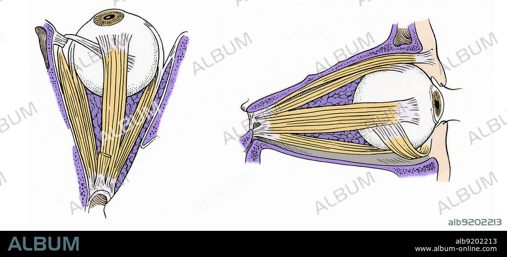
Illustration of Eye Muscles

|
Add to another lightbox |
|
Add to another lightbox |


Buy this image.
Select the use:

Title:
Illustration of Eye Muscles
Caption:
Anatomical illustration of the muscles of the eye in superior view (on left) and left lateral view (on right). The superior view shows the medial rectus, superior oblique, superior rectus, lateral rectus, and levator palprebrae superioris. The left lateral view shows the levator palpebrae, the superior rectus, the inferior rectus, the inferior oblique and the frontal sinus.
Credit:
Album / Science Source
Releases:
Image size:
5220 x 2379 px | 35.5 MB
Print size:
44.2 x 20.1 cm | 17.4 x 7.9 in (300 dpi)
Keywords:
ANATOMY • ART • BODY • BT7929 • COLOR • COLORIZED • ENHANCE • ENHANCED • EYE • EYEBALL • EYEBALLS • EYES • FRONT • FRONTAL • GROSS ANATOMY • HEALTHY • HUMAN • HUMANE • ILLUSTRATION • ILLUSTRATIONS • ILUSTRATION • INDIVIDUAL • INFERIOR • LATÉRAL • LATERALIS • LEFT • LEVATOR • MEDIAL • MUSCLE • MUSCLES • NORMAL • OBLIQUE • OPTICAL • PALPEBRAE • PALPREBRAE • PERSON • RECTUS • RF • SCIENCE • SIDE • SINUS • STRUCTURE • SUPERIOR • SUPERIORIS • MUSCLES


 Pinterest
 Twitter
 Facebook
 Copy link
 Email
Email
