alb10708043
MRI of Normal Sacrum
| Share |
|---|
 Pinterest Pinterest |
 Twitter Twitter |
 Facebook Facebook |
 Copy link Copy link |
 Email Email |
|
Add to another lightbox |
|
Add to another lightbox |


Title:
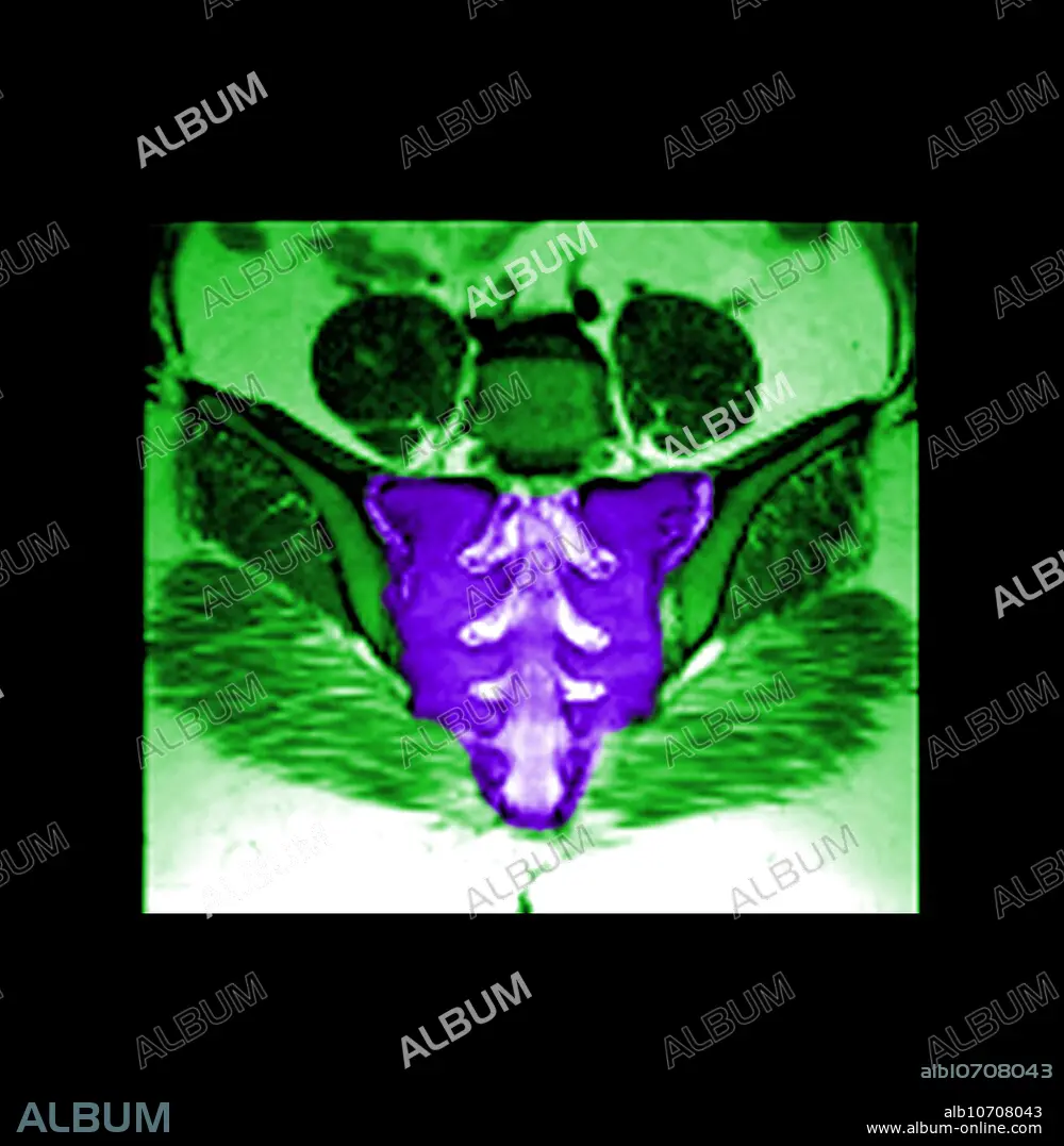
MRI of Normal Sacrum
Caption:
This color enhanced coronal (frontal) MRI image of the sacrum shows the normal anatomy of the sacrum, the sacroiliac joints, and the sacral neural foramina. MRI is an excellent tool to evaluate the sacrum, for most pathology.
Credit:
Album / Science Source / MEDICAL BODY SCANS
Releases:
Model: No - Property: No
Rights questions?
Rights questions?
Image size:
4172 x 4272 px | 51.0 MB
Print size:
35.3 x 36.2 cm | 13.9 x 14.2 in (300 dpi)
