alb10622680
Epigastric region

|
Add to another lightbox |
|
Add to another lightbox |


Title:
Epigastric region
Caption:
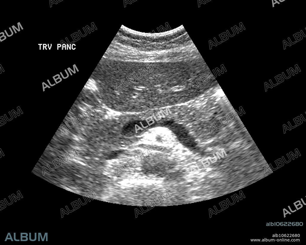
This axial (cross section) ultrasound image of the epigastric region shows a nice example of the normal appearance of the pancreas anterior (towards the top) to the splenic vein. Near the top of the image is the left lobe of the liver.
Credit:
Album / Science Source / LIVING ART ENTERPRISES, LLC
Releases:
Model: No - Property: No
Rights questions?
Rights questions?
Image size:
3600 x 2700 px | 27.8 MB
Print size:
30.5 x 22.9 cm | 12.0 x 9.0 in (300 dpi)
Keywords:
ABDOMEN • ABDOMINAL • ANATOMY • ANTERIOR • ANTERIORLY • AXIAL • BELLY • BODY • CROSS • DIGESTION • DIGESTIVE • ECHOGRAPHY • EPIGASTRIC • EPIGASTRIUM • GROSS ANATOMY • GUT • HEALTHY • HUMAN • HUMANE • IMAGE • IMAGING • INDIVIDUAL • LEFT • LIVER • LOBE • LOBULE • MEDICAL • MEDICINAL • NORMAL • OF • PANCREAS • PANCREATIC • PERSON • REGION • SCIENCE • SECTION • SECTIONAL • SONOGRAPHY • SPLENIC • STOMACH • SYSTEM • THE • ULTRA-SOUND • ULTRASOUND • UPPER • VEIN • VENA
 Pinterest
 Twitter
 Facebook
 Copy link
 Email
Email

