alb10664731
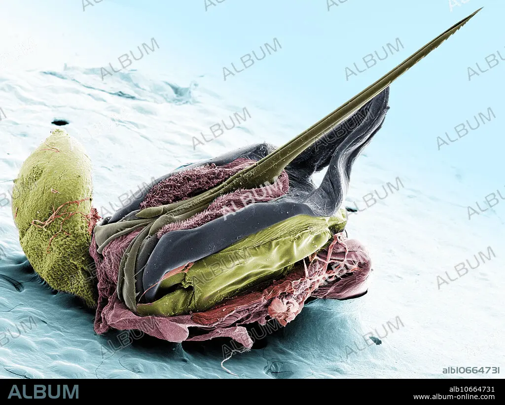
Honey bee stinger, SEM
| Share |
|---|
 Pinterest Pinterest |
 Twitter Twitter |
 Facebook Facebook |
 Copy link Copy link |
 Email Email |
|
Add to another lightbox |
|
Add to another lightbox |


Title:
Honey bee stinger, SEM
Caption:
Scanning electron microscope image of a Honey Bee Stinger, (Apis mellifera). The large sack on the left produces the poison. Once the barbs are set into the victim, the poison flows between the two blades of the stinger. Magnification: x50 when printed at 10 centimetres wide.
Credit:
Album / Science Source / TED KINSMAN
Releases:
Model: No - Property: No
Rights questions?
Rights questions?
Image size:
3600 x 2700 px | 27.8 MB
Print size:
30.5 x 22.9 cm | 12.0 x 9.0 in (300 dpi)
