alb10687860
Normal coronal and cross-sections of brain, illustration
| Share |
|---|
 Pinterest Pinterest |
 Twitter Twitter |
 Facebook Facebook |
 Copy link Copy link |
 Email Email |
|
Add to another lightbox |
|
Add to another lightbox |


Title:
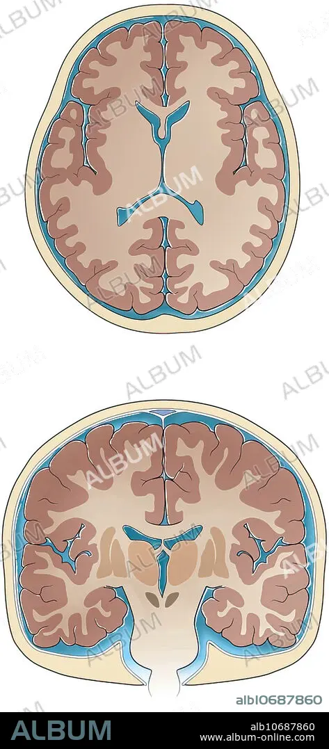
Normal coronal and cross-sections of brain, illustration
Caption:
Normal coronal and cross-sections of brain, illustration. Slices through the normal brain and skull showing ventricles, white matter, grey matter, basal ganglia, thalamus sulci and gyri, cerebrospinal fluid and skull.
Credit:
Album / Science Source / Sue Seif
Releases:
Model: No - Property: No
Rights questions?
Rights questions?
Image size:
2841 x 6150 px | 50.0 MB
Print size:
24.1 x 52.1 cm | 9.5 x 20.5 in (300 dpi)
Keywords:
ANATOMICAL • ANATOMY • ANATOMY: SKULL • ARTWORK • BASAL • BRAIN • BRAINS • CEREBROSPINAL • CORONAL • CRANEO • CRANEOS • CRANIUM • CRANIUMS • CROSS CUT • CROSS • CROSS-CUT • CROSS-SECTION • CROSS-SECTIONS • DRAWING • DRAWINGS • FLUID • GANGLIA • GREY • GROSS ANATOMY • GYRI • ILLUSTRATION • MATTER • NO ONE • NO-ONE • NOBODY • NORMAL • SECTION • SECTIONAL • SECTIONS • SKULL • SKULL, ANATOMY • SKULLS • SULCI • THALAMUS • VENTRICLES • WHITE
