alb3793110
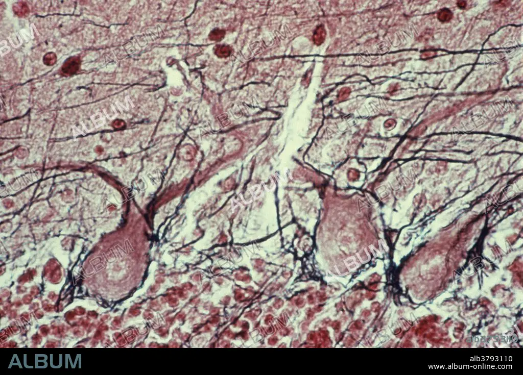
Purkinje Cells in the Cerebellum
| Share |
|---|
 Pinterest Pinterest |
 Twitter Twitter |
 Facebook Facebook |
 Copy link Copy link |
 Email Email |
|
Add to another lightbox |
|
Add to another lightbox |


Title:
Purkinje Cells in the Cerebellum
Caption:
Purkinje cells in the cerebellum. Axons appear black. The layers of the cerebellum consist of the "molecular layer" (outer), "granule cell layer" (inner), and an intervening layer of huge neurons called "Purkinje cells" H & E stain.
Credit:
Album / Science Source / Biophoto Associates
Releases:
Model: No - Property: No
Rights questions?
Rights questions?
Image size:
3278 x 2184 px | 20.5 MB
Print size:
27.8 x 18.5 cm | 10.9 x 7.3 in (300 dpi)
Keywords:
BRAIN • CELL • CEREBELLUM • GRANULE CELL • INTERVENING • LAYER • MOLECULAR • NEURAL • NEURON • PURKINJE
