alb10620608
ARACHNOID CYST
| Share |
|---|
 Pinterest Pinterest |
 Twitter Twitter |
 Facebook Facebook |
 Copy link Copy link |
 Email Email |
|
Add to another lightbox |
|
Add to another lightbox |


Title:
ARACHNOID CYST
Caption:
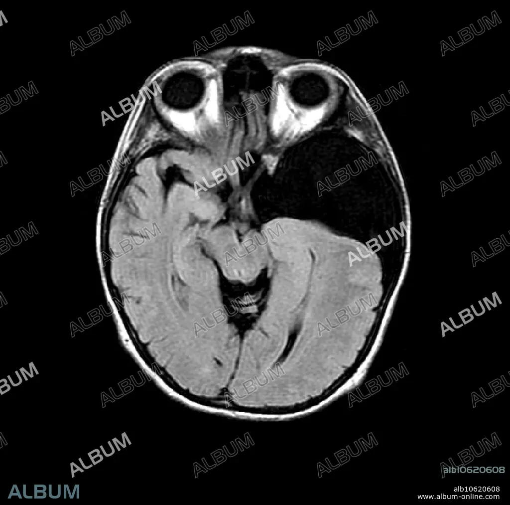
This axial (cross section) MRI image of the brain shows a large left middle cranial fossa arachnoid cyst (at right, dark). This is an abnormal collection of CSF (cerebral spinal fluid) which is walled off and acts like a mass. It is benign but can cause a variety of symptoms including seizures.
Credit:
Album / Science Source / LIVING ART ENTERPRISES, LLC
Releases:
Model: No - Property: No
Rights questions?
Rights questions?
Image size:
3842 x 3600 px | 39.6 MB
Print size:
32.5 x 30.5 cm | 12.8 x 12.0 in (300 dpi)
Keywords:
ABNORMAL • ANATOMY • ANATOMY: SKULL • ARACHNOID • AXIAL • BENIGN • BRAIN • CEREBRAL • CEREBRI • COLLECTION • CONDITION • CONFISCATION • CRANEO • CRANEOS • CRANIAL • CRANIUM • CRANIUMS • CROSS • CSF • CYST • DIAGNOSE • DIAGNOSIS • DIAGNOSTIC • DISEASE • DISORDER • FLUID • FOSSA • GROSS ANATOMY • HEALTHCARE • IMAGE • IMAGING • MAGNETIC • MEDICAL • MEDICINAL • MEDICINE • MRI • OF • REQUISITION • RESONANCE • SCIENCE • SECTION • SECTIONAL • SEIZURE • SKULL • SKULL, ANATOMY • SKULLS • SONORITY • SPINAL • THE
