alb9567838
Anatomy of Human Tongue, Illustration
| Share |
|---|
 Pinterest Pinterest |
 Twitter Twitter |
 Facebook Facebook |
 Copy link Copy link |
 Email Email |
|
Add to another lightbox |
|
Add to another lightbox |


Title:
Anatomy of Human Tongue, Illustration
Caption:
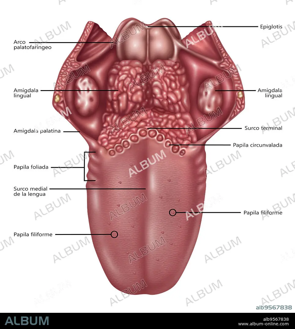
Illustration depicting the anatomy of the human tongue. Listed at left from top to bottom are: palatopharyngeal arch, lingual tonsil, palatoglossal arch, foliate papillae, medial sulcus of the tongue, filiform papilla. Listed at right from top to bottom are: epiglottis, palatine tonsil, terminal sulcus, vallate papilla (circumvallate papilla), fungiform papilla.
Credit:
Album / Science Source / Gwen Shockey
Releases:
Model: No - Property: No
Rights questions?
Rights questions?
Image size:
3300 x 3498 px | 33.0 MB
Print size:
27.9 x 29.6 cm | 11.0 x 11.7 in (300 dpi)
Keywords:
ANATOMICAL • ANATOMY • ANNOTATED • ART • ARTWORK • CIRCUMVALLATE PAPILLA • DRAWING • EPIGLOTTIS • FILIFORM PAPILLA • FOLIATE PAPILLAE • FUNGIFORM PAPILLA • GRAPHIC • GROSS ANATOMY • HUMAN TONGUE • ILLUSTRATION • ILLUSTRATIONS • INFOGRAPHIC • INFORMATION GRAPHIC • LINGUAL TONSIL • MEDIAL SULCUS OF THE TONGUE • MEDICAL • MEDICINAL • PALATINE TONSIL • PALATOGLOSSAL ARCH • PALATOPHARYNGEAL ARCH • PAPILLA • PAPILLAE • SCIENCE • SPANISH VERSION • TASTE BUDS • TERMINAL SULCUS • TONGUE ANATOMY • TONGUE • VALLATE PAPILLA • WHITE BACKGROUND
