alb10620762
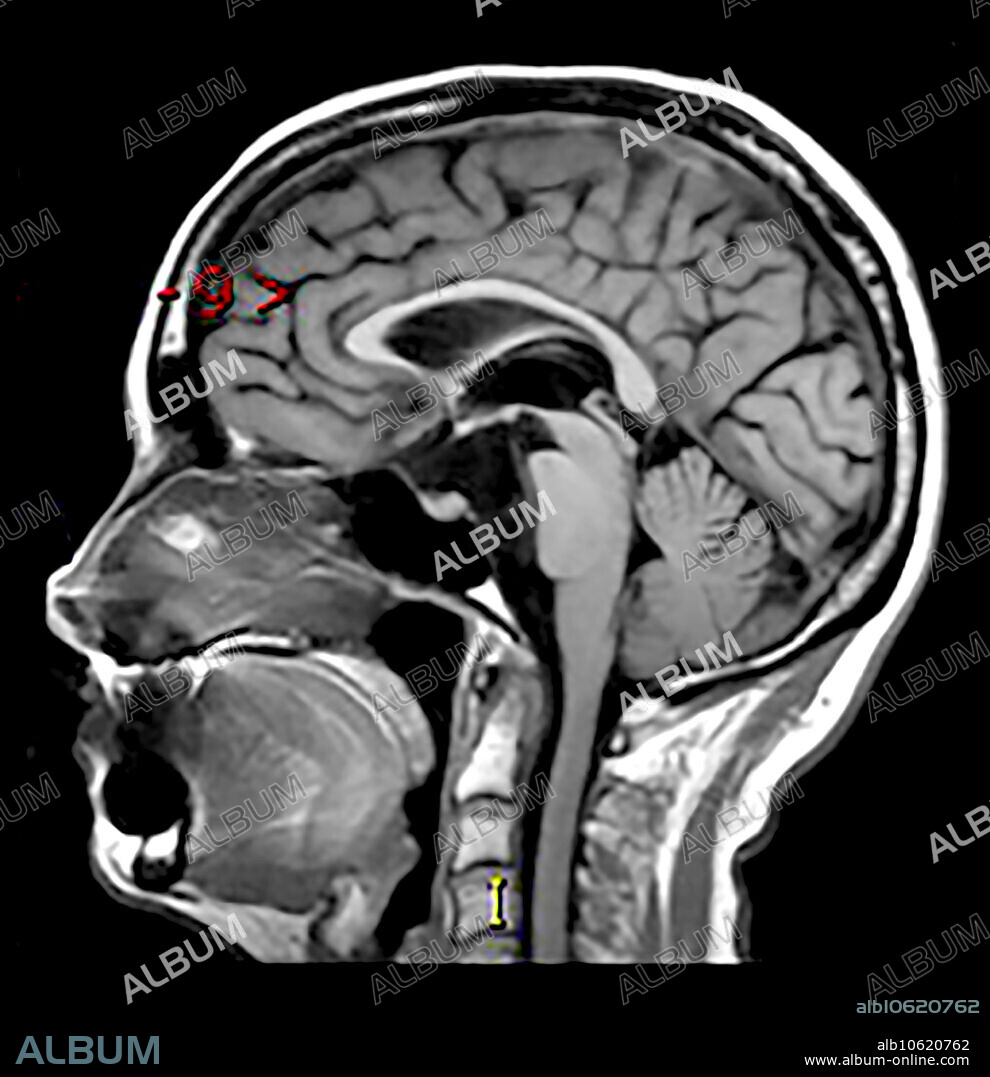
Epidermoid Tumour Prepontine Cistern

|
Add to another lightbox |
|
Add to another lightbox |


Title:
Epidermoid Tumour Prepontine Cistern
Caption:
This sagittal (from the side) T1 weighted MR image without contrast shows a hypointense mass like region in the prepontine cistern with compression of the brainstem. This represents an epidermoid tumour/cyst which is a congenital lesion consisting of keratinized epithelium with desquamation. These expand/grow over time. This is the same as a congenital cholesteatoma.
Credit:
Album / Living Art Enterprises, LLC/Science Source
Releases:
Model: No - Property: No
Rights questions?
Rights questions?
Image size:
4200 x 4346 px | 52.2 MB
Print size:
35.6 x 36.8 cm | 14.0 x 14.5 in (300 dpi)
Keywords:
ABNORMAL • BRAIN • BRAINSTEM • CHOLESTEATOMA • CISTERN • COMPRESSION • CONGENITAL • CYST • EPIDERMOID • FOSSA • HEAD • IMAGING • IN • INJURY • INTRACRANIAL • LESION • MAGNETIC • MEDICAL • MEDICINAL • MR • MRI • POSTERIOR • PREPONTINE • RESONANCE • SCAN • SONORITY • TUMOUR • WOUND
 Pinterest
 Twitter
 Facebook
 Copy link
 Email
Email
