alb10670462
MRI of Acoustic Schwannoma
| Share |
|---|
 Pinterest Pinterest |
 Twitter Twitter |
 Facebook Facebook |
 Copy link Copy link |
 Email Email |
|
Add to another lightbox |
|
Add to another lightbox |


Title:
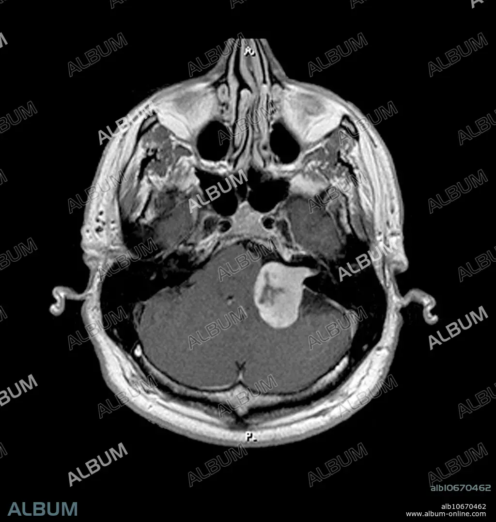
MRI of Acoustic Schwannoma
Caption:
This axial (cross sectional) MRI image of the brain at the level of the internal auditory canals shows a large enhancing tumour- in the left CP angle cistern and left internal auditory canal consistent with a large acoustic schwannoma. This caused unilateral hearing loss. MRI is the preferred method to evaluate patients with hearing loss.
Personalities:
Credit:
Album / Science Source / LIVING ART ENTERPRISES, LLC
Releases:
Model: No - Property: No
Rights questions?
Rights questions?
Image size:
3600 x 3600 px | 37.1 MB
Print size:
30.5 x 30.5 cm | 12.0 x 12.0 in (300 dpi)
Keywords:
ACOUSTIC • ACOUSTICS • ACUSTICA • AUDITORY • CANAL • CONDITION • DIAGNOSTIC • FOSSA • HEALTHCARE • HEARING • IMAGE • IMAGING • INJURY • INTERNAL • LESION • LOSS • MAGNETIC • MASS • MEDICAL • MEDICINAL • MEDICINE • MRI • NEUROMA • OF • PICTURE • POSTERIOR • RESONANCE • SCHWANNOMA • SONORITY • UNILATERAL • WOUND
