alb10670205
Massive Intracranial Hemorrhage, CT Scan
| Share |
|---|
 Pinterest Pinterest |
 Twitter Twitter |
 Facebook Facebook |
 Copy link Copy link |
 Email Email |
|
Add to another lightbox |
|
Add to another lightbox |


Title:
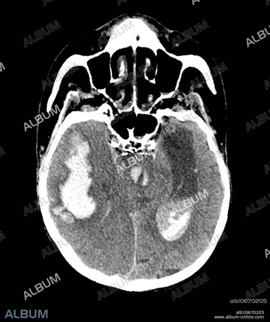
Massive Intracranial Hemorrhage, CT Scan
Caption:
This axial (cross-sectional) CT image of the brain in a hypertensive patient shows a large intracerebral hemorrhage (bleed) in the right temporal lobe with decompression/extension of the hemorrhage into the ventricular system. There is severe mass effect with brain herniation. The downward trans tentorial herniation results in tearing of perforating arteries arising from the basilar artery resulting in secondary brainstem hemorrhage (Duret Hemorrhage) seen in the central part of this image.
Personalities:
Credit:
Album / Science Source / Living Art Enterprises
Releases:
Model: No - Property: No
Rights questions?
Rights questions?
Image size:
4200 x 4763 px | 57.2 MB
Print size:
35.6 x 40.3 cm | 14.0 x 15.9 in (300 dpi)
Keywords:
ABNORMAL • ANIMAL: CAT • BLEED • BLEEDING • BLOOD • BRAIN • BRAINSTEM • CAT • CEREBRAL • CEREBRI • CONDITION • CRANIAL • CT • CVA • DIAGNOSTIC • DISEASE • DISORDER • DURET • FELIS CATUS • HAEMORRHAGIC • HEAD • HEMMORHAGE • HEMORRHAGE • HEMORRHAGED • HEMORRHAGING • HERNIA • HERNIATED • HERNIATION • HYPERTENSION • HYPERTENSIVE • IMAGING • INFARCTION • INJURY • INTESTINAL HERNIA • INTRACEREBRAL • INTRACRANIAL • INTRAVENTRICULAR • LESION • MASS • MEDICAL • MEDICINAL • MEDICINE • NEUROIMAGING • PATHOLOGY • SANGUINE • SCAN • SCIENCE • STROKE • UNHEALTHY • WOUND
