alb10611083
Multiple sclerosis, MRI

|
Add to another lightbox |
|
Add to another lightbox |


Title:
Multiple sclerosis, MRI
Caption:
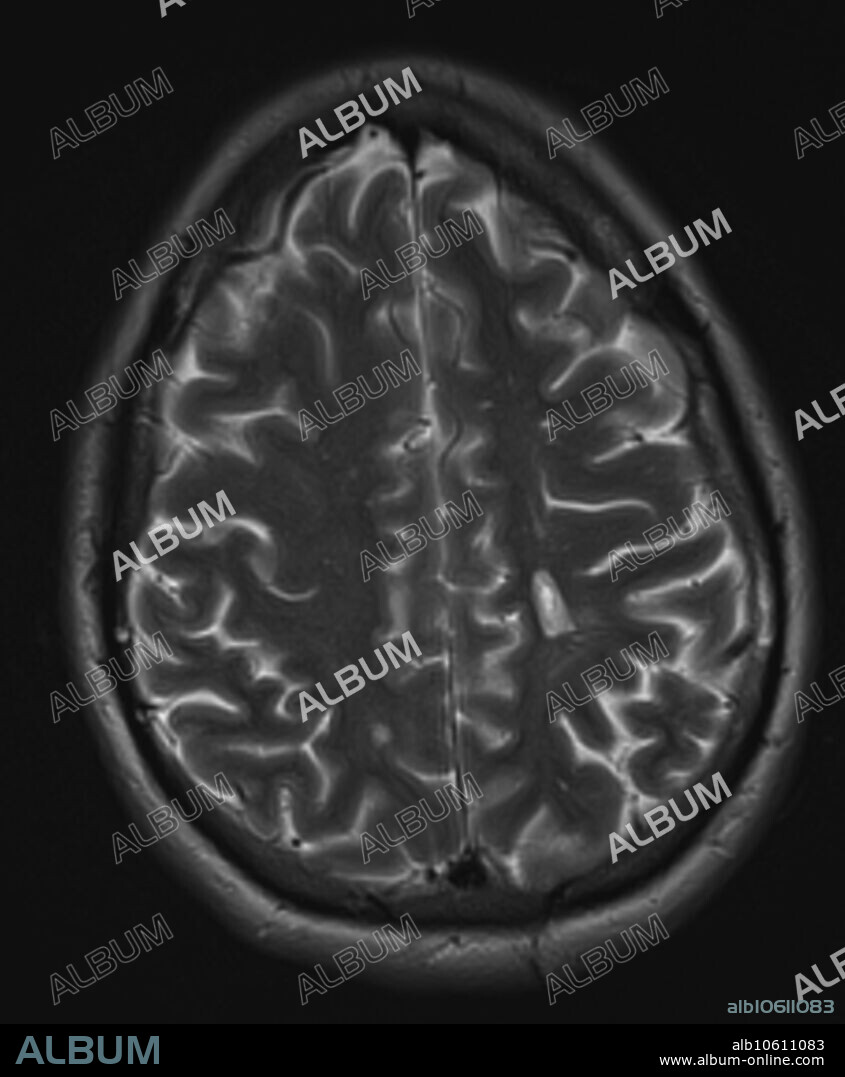
3.0T transverse, axial T2 MRI of 30 year old female with multiple sclerosis. Images reveal multiple foci of demyelinating disease throughout the periventricular and deep white matter. A chronic plaque results in a hole in the left parietal white matter centrum semiovale corona radiata.
Credit:
Album / Science Source / Steven Needell
Releases:
Model: No - Property: No
Rights questions?
Rights questions?
Image size:
1746 x 2117 px | 10.6 MB
Print size:
14.8 x 17.9 cm | 5.8 x 7.1 in (300 dpi)
Keywords:
3 • ABNORMAL • ACTIVE • BRAIN • CONDITION • CROSS-SECTIONAL • DARK • DAWSON • DEMYELINATING • DIAGNOSTIC • DISEASE • DISORDER • ENHANCEMENT • FEMALE • FINGERS • FLAIR • HOLE • IMAGING • MAGNETIC • MATTER • MEDICAL • MEDICINAL • MEDICINE • MRI • MS • MULTIPLE • PATHOLOGICAL • PATHOLOGY • PERIVENTRICULAR • PLAQUE • PLATE • RADIOLOGY • RESONANCE • SCAN • SCLEROSIS • SONORITY • T • T1 • T2 • TRANSVERSE • UNHEALTHY • WHITE
 Pinterest
 Twitter
 Facebook
 Copy link
 Email
Email

