alb3786579
Structure of Taste Bud, Illustration
| Share |
|---|
 Pinterest Pinterest |
 Twitter Twitter |
 Facebook Facebook |
 Copy link Copy link |
 Email Email |
|
Add to another lightbox |
|
Add to another lightbox |


Title:
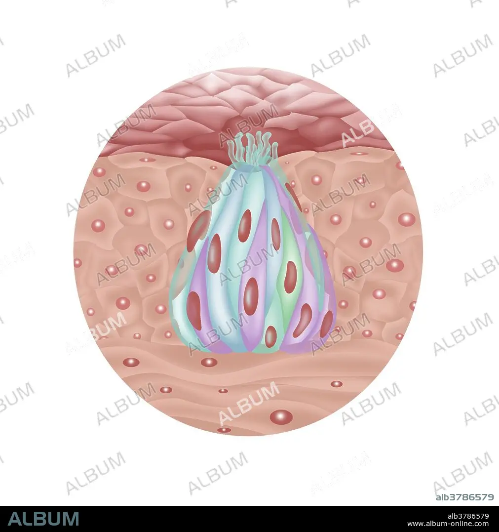
Structure of Taste Bud, Illustration
Caption:
Illustration of the structure of a taste bud. From top to bottom: Taste pore (dark pink area), gustatory hair (light green follicles), gustatory receptor cell (green & blue panels in pod), basal cell (reddish spots), stratified squamous epithelium (outer light pink area), supporting cell (purple areas), connective tissue (light pink area at bottom), sensory neurons.
Credit:
Album / Science Source / Gwen Shockey
Releases:
Model: No - Property: No
Rights questions?
Rights questions?
Image size:
1920 x 1944 px | 10.7 MB
Print size:
16.3 x 16.5 cm | 6.4 x 6.5 in (300 dpi)
Keywords:
