alb10623375
Mesial Parietal Meningioma MRI
| Share |
|---|
 Pinterest Pinterest |
 Twitter Twitter |
 Facebook Facebook |
 Copy link Copy link |
 Email Email |
|
Add to another lightbox |
|
Add to another lightbox |


Title:
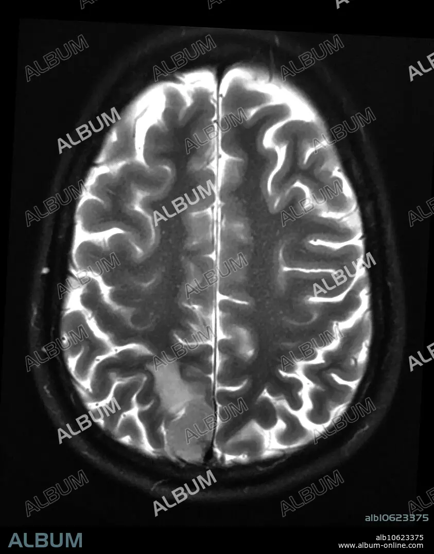
Mesial Parietal Meningioma MRI
Caption:
This axial (cross sectional) T2 weighted MR image shows a meningioma in the posterior-mesial aspect of the parietal lobe (also called the precuneus). Meningiomas are generally benign, non-aggressive tumours but can sometimes become malignant/aggressive.
Personalities:
Credit:
Album / Living Art Enterprises, LLC/Science Source
Releases:
Model: No - Property: No
Rights questions?
Rights questions?
Image size:
3900 x 4738 px | 52.9 MB
Print size:
33.0 x 40.1 cm | 13.0 x 15.8 in (300 dpi)
