alb10656953
Lamina Externa, TEM
| Share |
|---|
 Pinterest Pinterest |
 Twitter Twitter |
 Facebook Facebook |
 Copy link Copy link |
 Email Email |
|
Add to another lightbox |
|
Add to another lightbox |


Title:
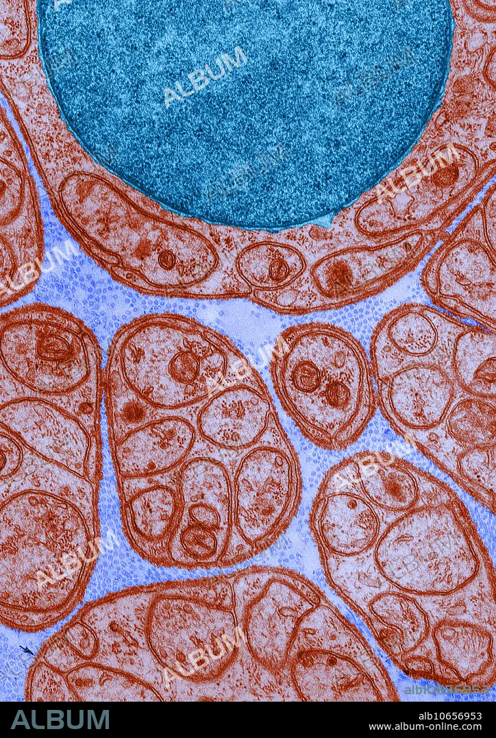
Lamina Externa, TEM
Caption:
Color enhanced transmission electron micrograph of a nerve in cross-section, showing groups of unmyelinated axons enclosed in deeply invaginated recesses in the surface of the Schwann cells. Around each Schwann cell is a thin, continuous lamina externa, especially visible at the arrows.
Credit:
Album / Science Source / DON W. FAWCETT
Releases:
Model: No - Property: No
Rights questions?
Rights questions?
Image size:
3490 x 4939 px | 49.3 MB
Print size:
29.5 x 41.8 cm | 11.6 x 16.5 in (300 dpi)
Keywords:
AXON • BASEMENT • BOUNDARY • CELL • CELLULAR • DENSA • ELECTRON • EXTERNA • FRONTIER • HISTOLOGY • LAMINA • LAYER • LIMIT • MEMBRANE • MESAXON • MESENTERY • MICROGRAPH • MICROGRAPHY • MICROSCOPY • MYELIN • MYELINATED • NERVE • PERIPHERAL • SCHWANN • SCIENCE • TEM • TRANSMISSION • UNMYELINATED
