alb10670497
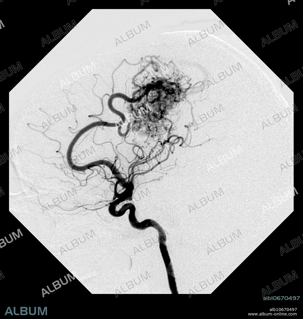
Cerebral Angiogram of AVM

|
Add to another lightbox |
|
Add to another lightbox |


Buy this image.
Select the use:

Title:
Cerebral Angiogram of AVM
Caption:
This lateral (from the side) image from an angiogram of the brains blood vessels shows the typical appearance of a high flow arterial venous malformation (AVM). There is enlargement of the pericallosal branch of the anterior cerebral artery supplying a large nidus (complex collection of abnormal blood vessels) and the very early appearance of the draining vein (along right side of the nidus). These are dangerous malformations of blood vessels that can lead to bleeding, stroke, seizures, headache and even death.
Credit:
Album / Science Source / Living Art Enterprises, LLC
Releases:
Image size:
4267 x 4267 px | 52.1 MB
Print size:
36.1 x 36.1 cm | 14.2 x 14.2 in (300 dpi)
Keywords:
ABNORMAL • ANGIOGRAM • ANGIOGRAPHY • ARTERIAL • ARTERIE • ARTERIES • AVM • BLEEDING • BLOOD • CEREBRAL • CEREBRI • CONGENITAL • FISH: RAY • FLOW • HEMMORHAGE • HEMORRHAGE • HEMORRHAGED • HEMORRHAGING • HIGH • IMAGE • INJURY • INTRACEREBRAL • INTRACRANIAL • LESION • MALFORMATION • OF • PICTURE • RAY • RAY, FISH • SANGUINE • VASCULAR • VEINOUS • VENOUS • VESSELS • WOUND • X • X-RAY • XRAY • ARTERIES


 Pinterest
 Twitter
 Facebook
 Copy link
 Email
Email
