alb3810639
Cork from Micrographia, 1665
| Share |
|---|
 Pinterest Pinterest |
 Twitter Twitter |
 Facebook Facebook |
 Copy link Copy link |
 Email Email |
|
Add to another lightbox |
|
Add to another lightbox |


Title:
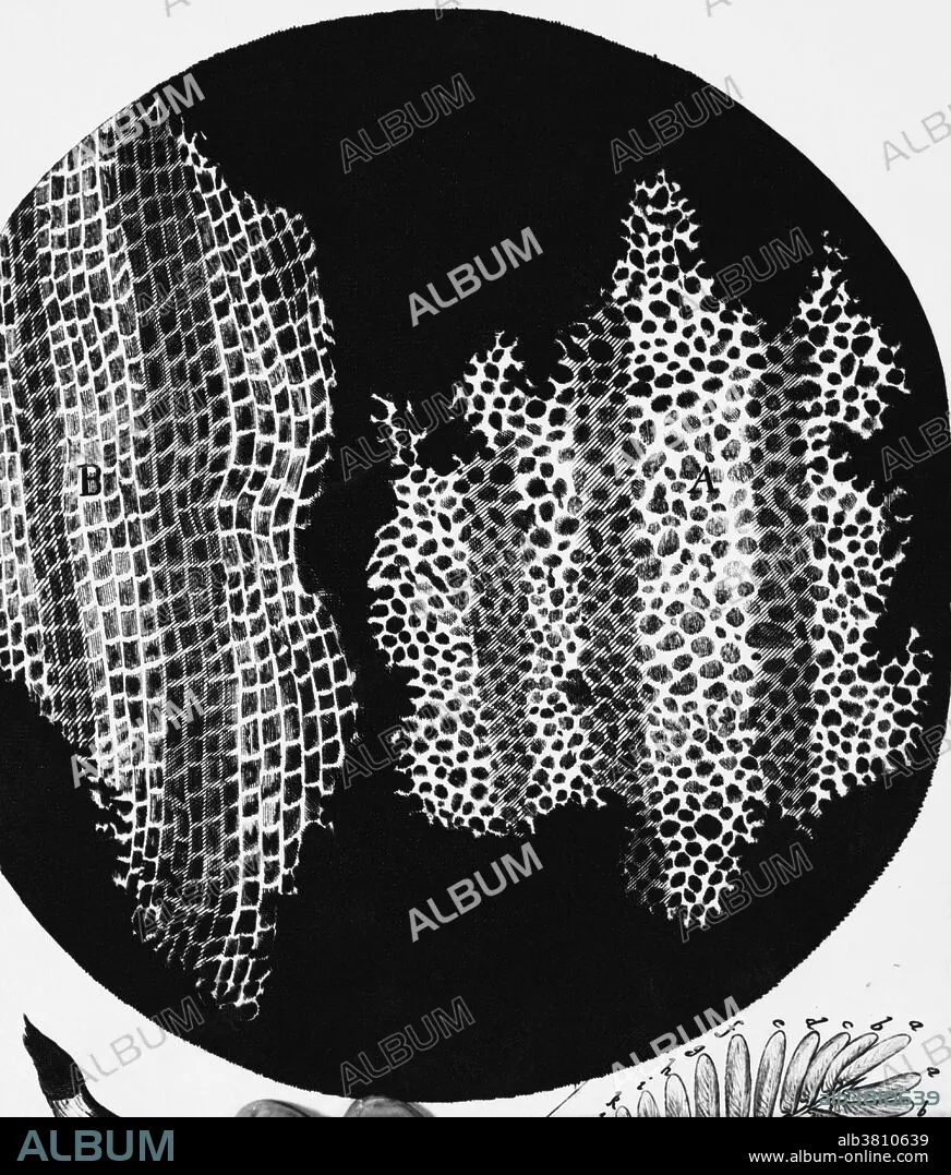
Cork from Micrographia, 1665
Caption:
Drawing of cork seen under a microscope; the first picture showing "cells" in a biological specimen, named as such by Robert Hooke. He prepared the specimen by making thin slices with a razor blade, and thus he invented the technique of sectioning; the discovery was described to the Royal Society of London on 13th April 1663. This figure first appeared in his book called "Micrographia"; this contains some of the most beautiful drawings of microscope observations ever made.
Credit:
Album / Science Source / Biophoto Associates
Releases:
Model: No - Property: No
Rights questions?
Rights questions?
Image size:
3858 x 4529 px | 50.0 MB
Print size:
32.7 x 38.3 cm | 12.9 x 15.1 in (300 dpi)
Keywords:
1665 • 17TH CENTURY • ART • ARTWORK • BLACK & WHITE • BLACK AND WHITE • BLACK BACKGROUND • BW • CELL • CELLULAR • CORK CELL • CORK CELLS • CORK • DIAGRAM • DRAWING • ENGRAVING • ETCHING • FAGACEAE • HISTORIC • HISTORICAL • HISTORY • HOOKE'S CORK CELLS • HOOKE • ILLUSTRATION • MICROGRAPHIA • MICROSCOPIC • MICROSCOPY • PLANT • PLANTA • PLANTAE • PRINT • QUERCUS SUBER • QUERCUS • ROBERT HOOKE • ROYAL SOCIETY • SEVENTEENTH CENTURY
