alb10687860
Normal coronal and cross-sections of brain, illustration
| Compartir |
|---|
 Pinterest Pinterest |
 Twitter Twitter |
 Facebook Facebook |
 Copiar enlace Copiar enlace |
 Email Email |
|
Añadir a otro lightbox |
|
Añadir a otro lightbox |


¿Ya tienes cuenta? Iniciar sesión
¿No tienes cuenta? Regístrate
Compra esta imagen

Título:
Normal coronal and cross-sections of brain, illustration
Descripción:
Ver traducción automática
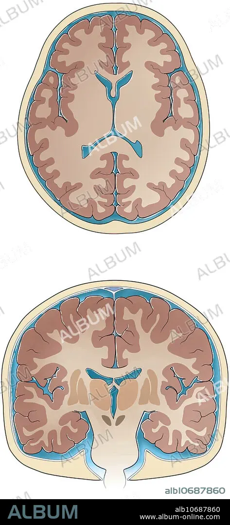
Normal coronal and cross-sections of brain, illustration. Slices through the normal brain and skull showing ventricles, white matter, grey matter, basal ganglia, thalamus sulci and gyri, cerebrospinal fluid and skull.
Crédito:
Album / Science Source / Sue Seif
Autorizaciones:
Modelo: No - Propiedad: No
¿Preguntas relacionadas con los derechos?
¿Preguntas relacionadas con los derechos?
Tamaño imagen:
2841 x 6150 px | 50.0 MB
Tamaño impresión:
24.1 x 52.1 cm | 9.5 x 20.5 in (300 dpi)
Palabras clave:
ANATOMIA • ANATOMICAS • BASALES • BLANCO • CALAVERA • CALAVERA. • CEFALORRAQUÍDEO • CEREBRO • CEREBROS • CIRCUNVOLUCIONES • CORTE TRANSVERSAL • CRANEO • CRANEOS • CRUZ • DIBUJO • DIBUJOS • GANGLIOS • GRIS • ILUSTRACION • NADIE • NORMALES • OBRA DE ARTE • SECCIÓN • SECCIONES • SECTION • SURCOS • TÁLAMO • TRAMO • VENTRICULOS
