alb10611083
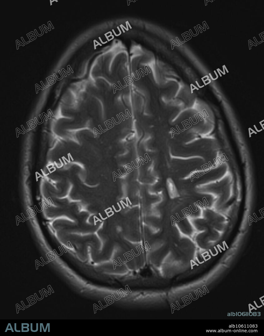
Multiple sclerosis, MRI

|
Añadir a otro lightbox |
|
Añadir a otro lightbox |


¿Ya tienes cuenta? Iniciar sesión
¿No tienes cuenta? Regístrate
Compra esta imagen

Título:
Multiple sclerosis, MRI
Descripción:
Ver traducción automática
3.0T transverse, axial T2 MRI of 30 year old female with multiple sclerosis. Images reveal multiple foci of demyelinating disease throughout the periventricular and deep white matter. A chronic plaque results in a hole in the left parietal white matter centrum semiovale corona radiata.
Crédito:
Album / Science Source / Steven Needell
Autorizaciones:
Modelo: No - Propiedad: No
¿Preguntas relacionadas con los derechos?
¿Preguntas relacionadas con los derechos?
Tamaño imagen:
1746 x 2117 px | 10.6 MB
Tamaño impresión:
14.8 x 17.9 cm | 5.8 x 7.1 in (300 dpi)
Palabras clave:
3 • ANORMAL • BLANCO • CEREBRO • CONDICION • DESORDEN • ENFERMEDAD • HOYO • IMAGENES • INSALUBRE • MEDICINA • MEDICINAL • MEJORA • MUJER • MULTIPLES • OSCURO • PATOLOGIA • PATOLÓGICOS • PLACA • PLACA, LA • RADIOLOGIA • RESONANCIA • RM • T1 • TRANSVERSAL
 Pinterest
 Twitter
 Facebook
 Copiar enlace
 Email
Email
