alb10679191
cervical spine
| Compartir |
|---|
 Pinterest Pinterest |
 Twitter Twitter |
 Facebook Facebook |
 Copiar enlace Copiar enlace |
 Email Email |
|
Añadir a otro lightbox |
|
Añadir a otro lightbox |


¿Ya tienes cuenta? Iniciar sesión
¿No tienes cuenta? Regístrate
Compra esta imagen

Título:
cervical spine
Descripción:
Ver traducción automática
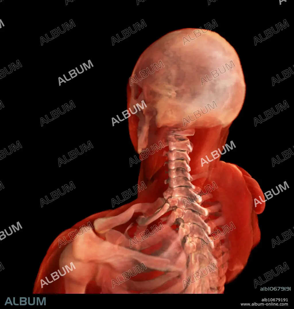
3D visualization based on scanned human data of posterolateral view of the cervical spine. The cervical spine consist of 7 vertebrae - C1-C7. The movements of the cervical spine are flexion and extension of the head which predominantly take place between the first cervical spine and the occipital bone of the skull. Rotation of the head occurs entirely at the joint between the first and second cervical vertebrae, the atlanto-axial joint.
Crédito:
Album / Science Source / ANATOMICAL TRAVELOGUE
Autorizaciones:
Modelo: No - Propiedad: No
¿Preguntas relacionadas con los derechos?
¿Preguntas relacionadas con los derechos?
Tamaño imagen:
2000 x 2000 px | 11.4 MB
Tamaño impresión:
16.9 x 16.9 cm | 6.7 x 6.7 in (300 dpi)
Palabras clave:
ACROMION • ANATOMIA • ANATOMICAS • ATLAS • CABEZA • CALAVERA • CALAVERA. • CIENCIA • CLAVÍCULA • COLUMNA • CRANEO • CRANEOS • CUELLO • CUERPO • DIMENSIONES • ESCÁPULA • ESCAPULARIO • ESPINAL • ESPINOSA • ESQUELETICO • HUESO • HUESOS • HUMERO • MEDICINA • MEDICINAL • PERSONA • PONER • PROCESO • PROCESOS • REGION • SALUD • SEGMENTO • SUPERIOR • TRES DIMENSIONES • TRES • VERTEBRALES
