alb10656953
Lamina Externa, TEM
| Compartir |
|---|
 Pinterest Pinterest |
 Twitter Twitter |
 Facebook Facebook |
 Copiar enlace Copiar enlace |
 Email Email |
|
Añadir a otro lightbox |
|
Añadir a otro lightbox |


¿Ya tienes cuenta? Iniciar sesión
¿No tienes cuenta? Regístrate
Compra esta imagen

Título:
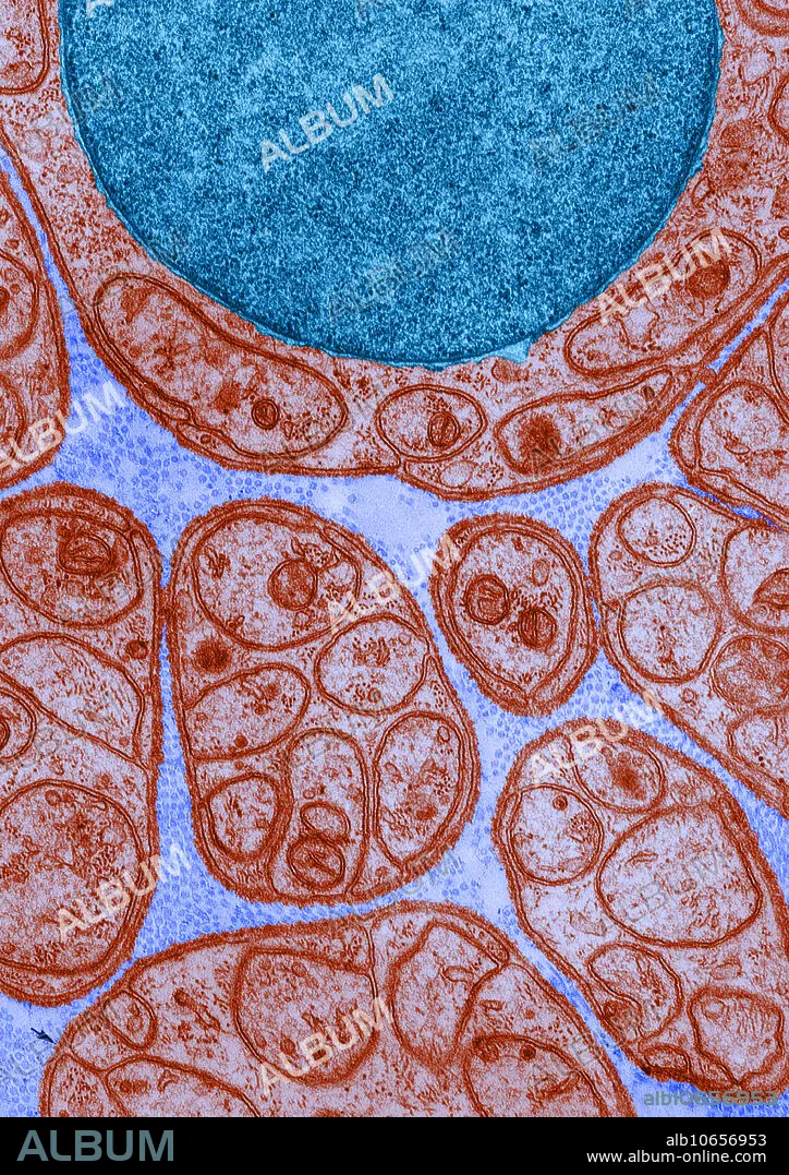
Lamina Externa, TEM
Descripción:
Ver traducción automática
Color enhanced transmission electron micrograph of a nerve in cross-section, showing groups of unmyelinated axons enclosed in deeply invaginated recesses in the surface of the Schwann cells. Around each Schwann cell is a thin, continuous lamina externa, especially visible at the arrows.
Crédito:
Album / Science Source / DON W. FAWCETT
Autorizaciones:
Modelo: No - Propiedad: No
¿Preguntas relacionadas con los derechos?
¿Preguntas relacionadas con los derechos?
Tamaño imagen:
3490 x 4939 px | 49.3 MB
Tamaño impresión:
29.5 x 41.8 cm | 11.6 x 16.5 in (300 dpi)
Palabras clave:
AXON • CELULA • CELULARES • CIENCIA • ELECTRON • LIMITE • MEMBRANA • MICROGRAFIA • MICROSCOPIA • MIELINA • NERVIO • PERIFÉRICOS • SCHWANN • TEMPERATURA • TRANSMISION
