alb10687860
Normal coronal and cross-sections of brain, illustration
| Partager |
|---|
 Pinterest Pinterest |
 Twitter Twitter |
 Facebook Facebook |
 Copier le lien Copier le lien |
 Email Email |
|
Ajouter à une autre Lightbox |
|
Ajouter à une autre Lightbox |


Avez-vous déjà un compte? S'identifier
Vous n'avez pas de compte ? S'inscrire
Acheter cette image

Titre:
Normal coronal and cross-sections of brain, illustration
Légende:
Voir la traduction automatique
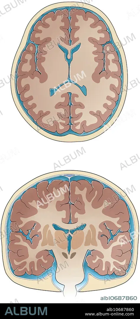
Normal coronal and cross-sections of brain, illustration. Slices through the normal brain and skull showing ventricles, white matter, grey matter, basal ganglia, thalamus sulci and gyri, cerebrospinal fluid and skull.
Crédit:
Album / Science Source / Sue Seif
Autorisations:
Modèle: Non - Propriété: Non
Questions sur les droits?
Questions sur les droits?
Taille de l'image:
2841 x 6150 px | 50.0 MB
Taille d'impression:
24.1 x 52.1 cm | 9.5 x 20.5 in (300 dpi)
Mots clés:
ANATOMIE • ANATOMIE: CRANE • ANATOMIE: CRANES • CORPS CRANE • CRANE • CRÂNES • GRIS • ILLUSTRATION • MORT CRANE • TETE DE MORT
