alb3788974
Top View of Normal Brain, Illustration
| Partager |
|---|
 Pinterest Pinterest |
 Twitter Twitter |
 Facebook Facebook |
 Copier le lien Copier le lien |
 Email Email |
|
Ajouter à une autre Lightbox |
|
Ajouter à une autre Lightbox |


Avez-vous déjà un compte? S'identifier
Vous n'avez pas de compte ? S'inscrire
Acheter cette image

Titre:
Top View of Normal Brain, Illustration
Légende:
Voir la traduction automatique
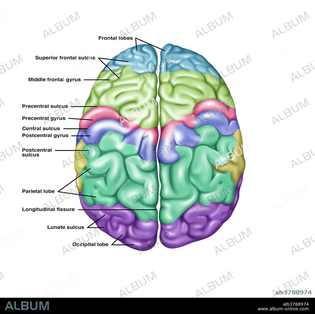
Illustration showing anatomy of a normal brain in a superior (top) view. Noted from top to bottom on the left side are the following: frontal lobes, superior frontal sulcus, middle frontal gyrus, precentral sulcus, precentral gyrus, central sulcus, postcentral gyrus, postcentral sulcus, parietal lobe, longitudinal fissure, lunate sulcus, and the occipital lobe.
Crédit:
Album / Science Source / Gwen Shockey
Autorisations:
Modèle: Non - Propriété: Non
Questions sur les droits?
Questions sur les droits?
Taille de l'image:
4337 x 4110 px | 51.0 MB
Taille d'impression:
36.7 x 34.8 cm | 14.5 x 13.7 in (300 dpi)
Mots clés:
