alb10679191
cervical spine
| Partager |
|---|
 Pinterest Pinterest |
 Twitter Twitter |
 Facebook Facebook |
 Copier le lien Copier le lien |
 Email Email |
|
Ajouter à une autre Lightbox |
|
Ajouter à une autre Lightbox |


Avez-vous déjà un compte? S'identifier
Vous n'avez pas de compte ? S'inscrire
Acheter cette image

Titre:
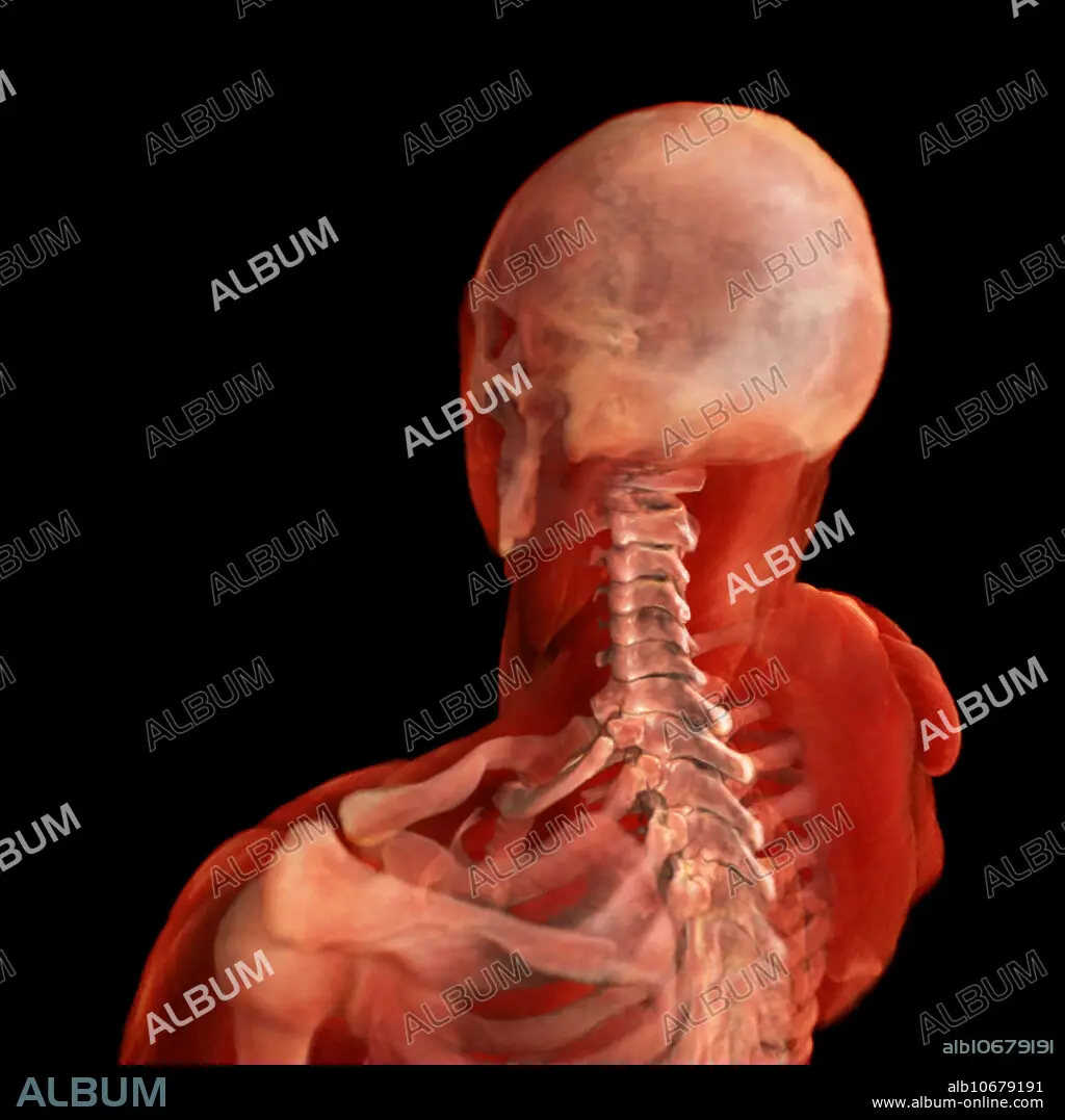
cervical spine
Légende:
Voir la traduction automatique
3D visualization based on scanned human data of posterolateral view of the cervical spine. The cervical spine consist of 7 vertebrae - C1-C7. The movements of the cervical spine are flexion and extension of the head which predominantly take place between the first cervical spine and the occipital bone of the skull. Rotation of the head occurs entirely at the joint between the first and second cervical vertebrae, the atlanto-axial joint.
Crédit:
Album / Science Source / ANATOMICAL TRAVELOGUE
Autorisations:
Modèle: Non - Propriété: Non
Questions sur les droits?
Questions sur les droits?
Taille de l'image:
2000 x 2000 px | 11.4 MB
Taille d'impression:
16.9 x 16.9 cm | 6.7 x 6.7 in (300 dpi)
Mots clés:
ANATOMIE • ANATOMIE: CRANE • ANATOMIE: CRANES • ANATOMIE: OS • CLAVICULE • CORPS CRANE • CORPS OS • COU • CRANE • CRÂNES • HUMAIN • MEDICAL • MORT CRANE • NUQUE • OS • OSSEMENT • PERSONNE • SCAPULAIRE • TERROIR • TETE DE MORT • TROIS
