alb10656953
Lamina Externa, TEM
| Partager |
|---|
 Pinterest Pinterest |
 Twitter Twitter |
 Facebook Facebook |
 Copier le lien Copier le lien |
 Email Email |
|
Ajouter à une autre Lightbox |
|
Ajouter à une autre Lightbox |


Avez-vous déjà un compte? S'identifier
Vous n'avez pas de compte ? S'inscrire
Acheter cette image

Titre:
Lamina Externa, TEM
Légende:
Voir la traduction automatique
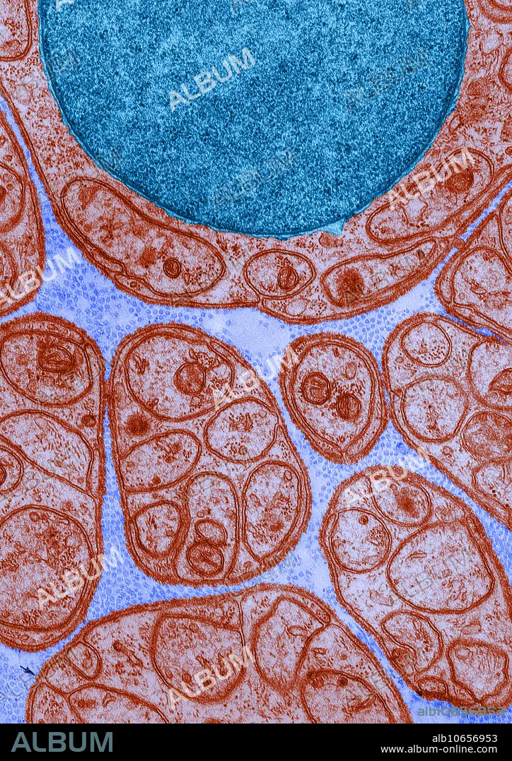
Color enhanced transmission electron micrograph of a nerve in cross-section, showing groups of unmyelinated axons enclosed in deeply invaginated recesses in the surface of the Schwann cells. Around each Schwann cell is a thin, continuous lamina externa, especially visible at the arrows.
Crédit:
Album / Science Source / DON W. FAWCETT
Autorisations:
Modèle: Non - Propriété: Non
Questions sur les droits?
Questions sur les droits?
Taille de l'image:
3490 x 4939 px | 49.3 MB
Taille d'impression:
29.5 x 41.8 cm | 11.6 x 16.5 in (300 dpi)
Mots clés:
